Tuesday, May 22, 2012

summary of osmoregulation

SUMMARY
The sand used for the building purpose contains  a lot of impurities like stones, shells ,mud particles and decayed particles of living things. The workers commonly use sieves as the apparatus for the removal  of  these waste materials ,like wise the living organism also produces waste material (nitrogenous substances, ions ,CO2 and water) as a by-product of various metabolic activities in the cell. Living organism also have an apparatus like sieve called excretory organ. They are responsible for the process of excretion or elimination of waste materials in the body.


Here we are mainly focused on the mechanism of elimination of nitrogenous waste materials.  The nitrogenous waste materials will be varies from organism to organism based on their habitat( where they located).Ammonia ,urea and uric acid are the different types of nitrogenous waste materials. Those organism which excretes
a)       Ammonia as the waste materials are called ammonotelic organism.eg:-Many bony fishes, Aq.amphibians and Aq. Insects.Ammonia is highly toxic in nature and their elimination require a large amount of water so  most of the ammonotelic  organism are seen in water.
b)       Urea as the waste materials are called ureotelic organism.eg:- Mammals,many terrestrial amphibians and marine fishes.urea is less  toxic than ammonia and it require  a less amount of water for their elimination .
c)       Uric acid as the waste materials are called uricotelic organism.eg:- Reptiles,birds ,landsnails and insects.
The excretory system in human being consist of a pair of kidney,a pair of ureters,a urinary bladder and a urethra. Each kidney has a millions of tubular structures called NEPHRONS.It is the basic unit of a kidney and has two portion.ie Glomerulus and renal tubule.Glomerulus is a group of fine capillaries formed from afferent arterioles .The renal tubule consist of double walled cup-like structure called Bowman’s capsule,proximal convoluted tubule(PCT),loop of Henle and distal convoluted tubule(DCT).The DCT of many nephrons joints to a common duct called collecting duct that ultimately open in to a renal pelvis through medullary pyramids.
Urine formation involves 3-process.ie,1) Glomerular filtration 2) Tubular reabsoreption 3)Tubular secretion.
The filtration is done by glomerulus due to high glomerular capillary blood preasure.about 1200ml of blood is filtered by the glomerulus per minute and forms 125ml of filtrate per minute in the bowman’s capsule, this is called glomerular filtration rate(GFR).JGA plays a significant role in  the regulation of the GFR.PCT is the major site of re-absorption and selective secretion.DCT and collecting  duct allows extensive re-absorption of water and certain electrolytes which helps in osmoregulation : H +K+ and NH3 could be secreted in to the filtrate by the tubule to maintain the ionic balance and pH of body fluid.
A counter current mechanism operates between the 2-limbs of the loop of Henle and vasa recta.The filtrate gets concentrated as it moves down the descending limb but is diluted by the ascending limb .Finally the urine is collected in the urinary bladder till a voluntary signal from CNS carries out its release through urethra. Skin ,lungs and liver. 

Other organs in Excretion

ROLE OF LIVER IN EXCRETION
The liver is a vital part of the excretory system, and the human body. It regulates glycogen storage, plasma protein synthesis, and drug detoxification. The liver secretes bile, a base used for breaking down fats. Therefore, it helps get rid of unneeded wastes in the body. It changes toxic ammonia, which is a poisonous gas , to urea, a harmless fluid. The kidney filters urea(a mix of sugar and waste) into a liquid called urine. The liver is the largest gland in the body.


ROLE OF LUNGS IN EXCRETION
Lungs are two-sponge like organs localized in each side of the thorax. They are constituted by pulmonary alveoli. They are not responsible for converting oxygen into carbon dioxide, but to maintain life supporting levels of these two gases in blood stream by excreting the extra carbon dioxide and keeping a regular supply of oxygen. Each and all organism's cells can take the oxygen through passive diffusion from the bloodstream and use it in its own metabolism, thus producing carbon dioxide that will be further excreted when passing through alveoli circulation.


ROLE OF SKIN IN EXCRETION
The skin is another part of the system, containing sweat that help regulate the concentration in one’s body while also keeping him or her cool. The salt helps evaporate the water, cooling off the skin. Sweat is excreted through sweat glands. There are two types of sweat glands: Eccrine sweat glands and Apocrine sweat glands. The basic purpose of skin is to provide a waterproof, protective, covering for the body's complex internal environment. The skin also plays a key role in helping to maintain the circulatory and nervous system.
ROLE OF ECCRINE GLANDS IN EXCRETION
The eccrine glands secrete mostly water and salt and are used by the body for temperature control. These glands are located all around the body but are most profuse around the soles of the feet, palms of the hands, and the forehead. They look like coiled tubes, spiraling towards the exterior of the skin
ROLE OF BILE IN EXCRETION
Bile is a greenish or yellowish substance secreted by the liver used for breaking down fats, ethanol, and acidic wastes. It is composed of water, cholesterol, lechithin, bicarbonate ions, bile salts, and pigments. Bile is a strong basic substance, classified as alkaline. It is released through bile ducts in the liver. A digestive chemical that is produce in the liver, stored in the gall bladder, and secreted in the small intestine

ROLE OF LARGE INTESTINE IN EXCRETION
The large intestine, or the colon, is the last part of the excretory system. It is the organ that removes solid waste from the body. Its function is not only to remove solid waste but to collect water from the waste that can be reused. It is part of the alimentary canal, a channel that flows through vertebrate animals. It is about 6 to 7 meters long and processes, transports, and excretes solid wastes.

MICTURITIONMICTURITION


MICTURITION
Micturition is  the process by which urine is expelled from the bladder.
The neural mechanism causing micturition is called Micturition reflex.

Micturition cycle occurs two phases .
 it consist of a filling phase and emptying phase.
Each phase requires a coordination interaction between the bladder and the nervous system.
Urine formed by the nephrone is ultimately carried to the urinary bladder.
Where it is stored till a voluntary signal is given by the central nervous system [CNS].
The signal is initiated by the stretching of the urinary bladder as it gets filled with urine.
In response ,the stretch receptors on the walls of the bladder send signals to the CNS.
The CNS passes on motor messages  to initiate the contraction of smooth muscles of the bladder .
The simultaneous relaxation of the urethral sphincter causing the release  of urine.
This type urine releasing process are called  MICTURITION

.
The urine formed is a light yellow coloured watery fluid .
The urine is a slightly acidic and has a characteristic odour.
An adult human excretes on average 1 to 1.5 litres of urine per day.
Analysis of  urine helps  in clinical diagnosis many metabolic disorders as well as malfunctioning of the kidney.
For Eg: Presence of glucose & ketone bodies in urine are indicative of diabetes mellitus.

Kidney functioning and regulating the volume and concentrating of body fluids

Kidney functioning and regulating the volume and concentrating of body fluids
The kidneys are responsible for keeping the homeostatically constant, which is achieved by regulating the volume and concentration of body fluids by selectively filtering and reabsorbing materials from the blood.
Blood volume control and kidney function
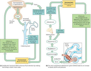
The volume of fluid in various fluid compartments in the body depends on the balance of fluid intake and fluid output. Fluid intake in response to thirst is a source of fluid, and receptor cells found in the hypothalamus activate thirst when exposed to hypertonic conditions - such as when water loss has been excessive or salt intake high.
Anti-diuretic hormone (ADH) and kidney function
ADH or antidiuretic hormone or vasopressin is the hormone produced from the hypothalamus of the brain. It is secreted into the blood from the posterior lobe of pituitary. ADH is secreted when the osmoreceptors of hypothalamus detect an increased osmolarity or lesser water concentration in the blood. This causes thirst, and the released ADH makes the distal convoluted tubule and collecting duct of nephrons permeable to water. The primary effect of ADH is to limit the amount of water being lost in urine,. It is done by increasing the amount of reabsorption of water into the blood. The ADH targets the cells of the tubules and collecting ducts, which causes an increase of permeability of the cell surfaces, where the water then leaves the renal tubules in to the blood by means of osmosis.
With more fluid being reabsorbed, the blood volume increases
Aldosterone hormone - regulation of sodium and potassium and kidney function
ADH is not the only hormone that helps with the regulation of kidney function - aldosterone (from the adrenal cortex) as well as parathyroid hormone (from the parathyroid glands) affects the balance and regulation of electrolyte content of the blood and body fluids.
When aldosterone is present in the blood, the distal renal tubules increase their re-absorption of sodium and the secretion of potassium. With this action, more water is retained in the body
Aldosterone is secreted by the adrenal glands when the level of the potassium in the blood is increased, as well as the self-regulatory action of the kidneys by means of the renin-angiotensin system.


Renin-angiotensin system regulation of kidney function
When blood pressure increases so does the glomerular filtration increase, but when blood pressure falls, the filtration level drops and the glomerular filtration rate then needs another system to increase the filtration rate.
This is made possible by a specialized region of the nephron - the juxtaglomerular complex
In mammalian nephrons the distal convoluted tubule forms a secretory tissue near the Bowman’s capsule, in between the afferent and efferent arteriole, called Juxtaglomerular apparatus (JGA).It has directed contact with the afferent arteriole. If the blood pressure or blood volume in the afferent arteriole is decreased, the JGA secretes an enzyme called renin into the blood stream. Renin converts and inactive plasma protein angiotensinogen to active angiotensin II. This angiotensin II behaves like a hormone and increase blood pressure and blood volume by the following mechanisms.
1.      It constricts arteries to reduce arterial volume, thereby increasing blood pressure.
2.      It stimulates the proximal convoluted part of the nephrons to reabsorb more water, Na+ and Cl-  ; which increase the blood volume.
3.      It stimulates the adrenal gland to release aldosterone, enhances the re-absorption of water and Na+ by the distal convoluted part of nephrons. This activity further increase the blood volume.
                  Thus the Renin- Angiotensin- Aldosterone system (RAAS) regulate the renal function to raise blood volume and blood pressure to a higher level.
                 Increased blood volume and blood pressure stimulates the auricular wall of heart to produse another protein called Atrial Natriuretic Factor (ANF). It inhibits the release of renin from JGA. Thus the balanced interplay between RAAS and ANF helps to maintain the blood pressure and blood volume normal

MECHANISM OF CONCENTRATION OF THE FILTERATE

MECHANISM  OF CONCENTRATION OF THE  FILTERATE


                    Mammals have the ability to produce a concentrated urine. The human body does not

       maintain a constant water volume, the kidneys use a transportsystem called the counter-

      current mechanism..The Henle’s loop and vasa recta play a significant role in this.

      The particles and fluid removed from the blood, the filtrate, moves from the Bowman's

    capsule to the proximal tubule, loops of Henle, distal tubule, and collecting tubule.

    The flow of filtrate in the two limbs of  henle’s loop is in opposite directions and thus forms a

    counter current The flow of blood through the limbs of vasa recta is also in a counter current

    pattern. The name is based on the fact that concentration first increases in the direction of

   f low, then decreases as flow continues through the ascending parallel loop. The mechanism

   relies on the adjacent, parallel loops of Henle and vasa recta.


In the ascending loop, Na+ (or any solute) is actively pumped out of the tubule. As flow  

   continues up the loop, the tubular concentration decreases as does the interstitial (the fluid

   surrounding the loop) concentration. Because water is impermeable in the ascending loop, the

   volume at the bottom of the loop is the same as that entering the distal tubule. At the bottom

   of the loop, the tubular and interstitial concentrations are equal.

In the descending loop, the concentrations inside and outside the tubule are increasing

   with the current, with the maximum concentration being reached at the bottom of the loop.

  The increased concentration is the result of the passive diffusion of Na+ into the tubule and

  water out of the tubule. When the filtrate reaches the distal tubule, a net loss of Na+ and water

  has occurred through the loops of  Henle.


Inside the distal and collecting tubules, the filtrate is either diluted or concentrated to

form urine. The Henle's loop and vasa recta are close together and counter current mechanism in them help 

in maintaining an osmolarity towards the tissue surrounded by Henles loop in the renal medulla.,.i.e.,from 

300 mOsmoIL in the cortex to about  1200 mOsmoIL in the inner medulla.This  gradient  is mainly caused 

by NaCl and urea.NaCl is transported by the ascending limb of Henle’loop which is exchanged with the 

descending limb of vasa recta.Nacl  is returned to the interstitium by the ascending portion of vasa 

recta.Similarly,small amount of urea enter the thin segment of the ascending limb of Henle’s loop which is 

transported back to the interstitium by the collecting tubule.

This counter current mechanism helps to maintain a concentration gradient in the medullary

interstitium.Presence of such interstitial gradient helps in an easy passage of water from the

collecting tubule thereby concentrating the filtrate(urine).human kidneys can produce urine nearly

four times  concentrated than the initial filtrate formed. 

urine formation

INTRODUCTION
Every one of us depends on the process of urination for the removal of certain waste products in the body. The production of urine is vital to the health of the body. We could not survive if we did not produce urine and eliminate it. Urine is composed of water, certain electrolytes, and various waste products that are filtered out of the blood system.. A major part of “cleaning" of the blood takes place in the kidneys and, particularly in the nephrons, where the blood is filtered to produce the urine. Both kidneys in the body carry out this essential blood cleansing function. Normally, about 20% of the total blood pumped by the heart each minute will enter the kidneys to undergo filtration. This is called the filtration fraction. The rest of the blood (about 80%) does not go through the filtering portion of the kidney, but flows through the rest of the body to service the various nutritional, respiratory, and other needs that are always present.

                            Urine formation involves three main processes namely, glomerular filtration, re absorption and secretion, that take place in different parts of the nephron
           

The three steps of urine formation are
A.    GLOMERULAR FILTRATION,

  1. TUBULAR REABSORPTION, and

  1. TUBULAR SECRETION.

o   GLOMERULAR FILTRATION occurs in the GLOMERULUS,
o   TUBULAR REABSORPTION occurs through the PROXIMAL CONVOLUTED TUBULE,
o   And TUBULAR SECRETION occurs through the DISTAL CONVOLUTED TUBULES.
o   GLOMERULAR FILTRATION is defined as the process of primary filtration taken place from the glomerulus in to the Bowmans capsule.
o   TUBULAR REABSORPTION is the process of reclaiming water and solutes from the tubular fluid and returning them in to the blood.
o   TUBULAR SECRETION is defined as the process of movement of  “wastes and excesses”  from the blood in to  the Distal and collecting tubule.
Filtration


Urine formation begins with the process of Glomerular filtration,. The water, waste products, salt, glucose, and other chemicals that have been filtered out of the blood are collectively known as glomerular filtrate. The glomerular filtrate consists primarily of water, excess salts (primarily Na+ and K+), glucose, and a waste product of the body called urea. Urea is formed in the body to eliminate the very toxic ammonia products that are formed in the liver from amino acids. Since humans cannot excrete ammonia, it is converted to the less dangerous urea and then filtered out of the blood. Urea is the most abundant of the waste products that must be excreted by the kidneys. The total rate of glomerular filtration (glomerular filtration rate or GFR) for the whole body  is normally about 125 ml per minute. That is, about 125 ml of water and dissolved substances are filtered out of the blood per minute. The following calculations may help you visualize how enormous this volume is. The GFR per hour is:
125 ml/min X 60min/hr= 7500 ml/hr.

 Reabsorption
The GFR per day is:
7500 ml/hr X 24 hr/day = 180,000 ml/day or 180 l
THE EXCRETORY SYSTEM


                         Just as how we thrown away the waste materials from our homes, our body to has a system to remove the waste. The system is called the excretory system. The excretory system consists  of  a pair of kidneys , ureters, the urinary bladder, a urethra. The main organs of the excretory system are the kidneys. There are two reddish brown bean shaped organs located on each side of the lower abdomen. Each kidney of an adult human measures 10-12 cm in length,5-7cm in width, 2-3cm in thickness with an average weight of 120-170 g . Towards the centre of the inner concave surface of the kidney is a notch called hilum through which ureter , blood vessels and nerves enter.    The kidneys filter the blood. They help to get rid of waste in the form of urine.  The urine is carried by tubes called ureters to the urinary bladder. The urinary bladder stores the urine. When the bladder is full, the urine is passed out through the urethra. In this way the excretory system removes the wastes from the body.

                    A detailed anatomy of the kidney showed here. The longitudinal section of the kidney shows the location of the renal cortex, renal medulla, the renal pyramids and the renal pelvis.
The outer layer of kidney is a tough capsule. Inside the kidney, there are two zones. An outer cortex  and inner medulla. The medulla is divided into a few conical masses projecting into the calyces. These conical masses are known as medullary pyramids. The cortex extends in between the medullary pyramids as renal columns called columns of bertini.
           Each kidney has nearly one million complex tubular structures called nephrons. Nephrons are the functional units and they are also known as kidney tubule.

              Observe the placement of the nephrons in the enlarged renal nerve. Seen here is the anatomy of nephron.

             The main parts of nephrons are the bowman’s capsule, the glomerulus, the proximal  convoluted tubule, the distal convoluted tubule and the loop of Henle.
              Glomerulus is a tuft of capillaries formed by the afferent arteriole. Blood from the glomerulus is carried away by an efferent arteriole.  The renal tubule begins with a double walled cup- like structure called bowman’s capsule . The bowman’s capsule encloses  the glomerulus.                   Glomerulus along with  bowman’s  capsule is called the malpighian body or renal corpuscle.(figure 4). The tubule continues further to form a highly coiled network called proximal convoluted tubule(PCT).A hairpin shaped Henle’s loop is the next part of the tubule which has a descending and an ascending limb. The ascending limb continues as another highly coiled tubular region called distal convoluted tubule(DCT). The DCTs of many nephrons open into a straight tube called collecting duct.

              The efferent arteriole emergingfrom the glomerulus forms a fine capillary network around the renal tubule called the peritubular capillaries. Aminute vessel of this network runsparallel to the Henle’s loop forming a ‘U’ shaped vasa recta.
              The structure of human excretory system, the kidneys and the nephrons can be observed here.


Tuesday, May 15, 2012

Introduction


INTRODUCTION
Excretion is the process by which waste products of metabolism and other non-useful materials are eliminated from an organismEvery organism, from the smallest protest to the largest mammal, must rid itself of the potentially harmful by-products of its own vital activities. This process in living things is called elimination.Example of toxic waste materials are ammonia, urea, uric acid , CO, H2O ,and ions like Na+, K+ ,Cl- ,phosphate ,sulphate, etc.Through excretion organisms control osmotic pressure - the balance between inorganic ions and water—and maintain acid-base balance. The process thus promotes homeostasis, the constancy of the organism’s internal environment
In single-celled organisms, waste products are discharged directly through the surface of the cell. Multicellular organisms utilize more complex excretory methods. Animals have special excretory organsand different excretory organs constitute the excretory system. . In man this is primarily carried out by the lungskidneys and skin.[1]

The main function of excretory system is to eliminate the nitrogen waste.In this chapter you will learn the mechanism of elimination of nitrogenous wastes. Ammonia, Urea,and Uric acid are the major forms of nitrogenous wastes. Ammonia is highly toxic and water soluble.The process of excreting ammonia is known asammonotelism.Such animals are called ammonotelic. It is found in aquatic animal groups like sponges, coelentrates, crustaceans, echinoderms, bony fish, tadpole larvae, and salamander.
Mammals, many terrestrial amphibians and marine fishes mainly excrete urea.They are called ureotelic.It is commonly found in man , whales, seals, desert mammals like kangaroo rats , camels, toads, frogs, cartilaginous fishes, aquatic and semi aquatic reptiles like alligator, terrapinsand turtles.Urea is far less toxic than ammonia. Ammonia produced by metabolism is converted into urea to the liver of these animals and released into the blood which is filtered and excreted out by the kidneys. To maintain a desired osmolarity,some urea may be retained in the kidney matrix of these animals.


Reptiles, birds, land snails, and insects excrete nitrogenous wastes as uric acid in the form of pellet or paste with a minimum loss of water. They are called uricotelic animals.Uric acid is formed from ammonia mostly in the liver and to some extent in kidneys. But itis much less toxic than both ammonia and urea and it is almost insoluble in water and can beeliminated  from the body in nearly a solid state, saving a lot of water.
Other Nitrogenous wastes –Allantosm, creatine, creatinine and hippouric acid are some nitrogenous wastes products excreted by mammals.
In most of the invertebrates, the excretory organs are simple tubular forms whereas in vertebrates , the excretory organs are complex.For example flate worms like Planeria have the simplest tubular excretory system called the Protonephridia or flame cells.


 Rotifers, some annelids and cephalochordaes (Amphioxus) also possess protonephrida.
Protonepridia are generally concerned with osmoregulation i.e., the regulation of ionic and fluid volume. Earthworms possess a similar simple tubular excretory system called metanephridia . 


\In cockroaches and other insects , the malpighian tubules are the excretory organs.


              Crustaceans like Cray fish have a pair of specialised excretory structures called ‘antennal gland’ or‘green gland’ that is situated at the base of the second antennae.